v
Stem Cell Engineering and Germplasm Resource Innovation

Research progress on pluripotent stem cell-derived organoids

  • Xueting Li , 1 ,
  • Jiannan Li 1 ,
  • Jinlian Hua , 1, 2
Expand
  • 1. College of Veterinary Medicine, Northwest A&F University, Yangling, 712100, Shaanxi
  • 2. Shaanxi Provincial Stem Cell Engineering Technology Research Center, Yangling, 712100, Shaanxi

Received date: 2025-12-31

  Online published: 2026-09-10

Abstract

Pluripotent stem cells are mainly derived from embryonic stem cells and induced pluripotent stem cells. Although embryonic stem cells have the potential of infinite proliferation and multi-directional differentiation, their application is restricted by ethical issues. In recent years, the deep integration of pluripotent stem cells and three-dimensional organoid technology has made in vitro model research officially enter the era of "quasi-physiology". This technology is the first to open a breakthrough in the field of human medicine. At present, it is providing a technical model for disease resistance breeding in the field of livestock and poultry through cross-species technology migration. In this context, this article focuses on the organoids derived from pluripotent stem cells. First, it focuses on reviewing key developmental signaling pathways such as Wnt, BMP, and TGF-β. Researchers need to differentially regulate the above pathways according to the types of organoids to be constructed. For example, when inducing neural induction of pluripotent stem cells into brain organoids, it is necessary to simultaneously block the BMP and TGF-β signaling pathways. In addition, this paper reviews the progress in the modeling of the pathological mechanisms of diseases in complex organs such as nerves, heart, and lungs, and discusses the frontier applications of various organs in the construction of infection and tumor models, drug screening, and toxicity assessment. It focuses on its key role in simulating disease processes, revealing pathogenic mechanisms, developing new therapies, and evaluating drug safety, and carefully reviews the innovative technical methods and progress of organoid culture systems in recent years. Combined with the current technical challenges faced by animal pluripotent stem cell-derived organs in model construction, it is proposed that the mature experience of human-derived organs in complex disease modeling should be used for reference. It provides a new research method and technical support for the sustainable development of livestock and poultry breeding in disease resistance breeding and excellent trait screening.

Cite this article

Xueting Li , Jiannan Li , Jinlian Hua . Research progress on pluripotent stem cell-derived organoids[J]. The Chinese Livestock and Poultry Breeding, 2026 , 22(9) : 85 -94 . DOI: 10.19543/j.cnki.1673-4556.20260629.001

多能干细胞(Pluripotent stem cells, PSCs)依据主要来源可分为两类:胚胎干细胞(Embryonic stem cell, ESC)与诱导多能干细胞(Induced pluripotent stem cell, iPSC)。胚胎干细胞源自早期胚胎,具有无限增殖和多向分化的能力,能分化成所有3个胚层的细胞,是疾病与再生医学研究的基石[1-2]。然而,其应用长期受伦理问题的制约[3]。这一瓶颈的突破源于2006年Takahashi和Yamanaka的开创性工作,他们通过导入特定转录因子,将体细胞重编程为诱导多能干细胞[4]。iPSC不仅拥有与ESC类似的自我更新和多能性,成功解决了伦理困境,并能提供几乎无限的、可追溯至特定个体(包括健康与疾病状态)的多能干细胞系,从而为精准医学研究开辟了全新途径[5-6]。
类器官技术标志着体外模型研究的一次革命性飞跃,与传统的二维培养相比,其核心优势在于能够通过三维(3D)培养体系模拟细胞在自然状态下的生长微环境[7]。该术语最初于1946年被用于描述类似于器官的畸胎瘤,但其现代定义则更为精确,特指由原代组织或多能干细胞在体外自组装形成的三维结构,这些结构能够模拟源器官的复杂细胞组成、空间构架及部分功能[1,8]。根据细胞来源,类器官主要可分为两大类:一是由成体干细胞衍生,例如2009年Sato等[1-2,4,9]利用“LGR5+肠道干细胞”培育出的首个上皮类器官,为该领域奠定了基础;二是由多能干细胞(包括胚胎干细胞和诱导多能干细胞)分化而来。
在众多来源中,多能干细胞源类器官展现出独特的巨大潜力。其被青睐的主要原因有3个:首先,iPSC可通过体细胞重编程获得,规避了ESC相关的伦理争议[5-6]。其次,iPSC技术使得从任何特定个体获取细胞成为可能,能够构建大规模、个性化的疾病模型。尤为重要的是,研究发现iPSC衍生的类器官在发育阶段上更接近于胎儿组织而非成人组织,这一特性使其成为研究胚胎发育事件和器官发生的优秀系统[10-11]。最后,iPSC具有无限增殖和多向分化的能力,能够构建那些难以从成体组织中获取干细胞的复杂器官模型,例如脑、肾等类器官仅被证明可由iPSC成功生成[12]。
本文聚焦于多能干细胞源类器官,探讨其核心培养技术与方案的最新优化,继而选取脑、肝、肾等代表性器官模型,综述其在模拟人类发育与疾病中的前沿研究进展,重点探讨 类器官在药物筛选、疾病建模及再生医学等转化研究中的新兴应用与未来挑战,旨在为该领域的后续研究提供系统性参考与创新性思路。

1 多能干细胞源类器官培养系统

将多能干细胞定向分化为类器官是一个精细调控的过程,该过程始于细胞在悬浮培养中聚集形成胚胎样体(Embryoid body, EB),随后通过添加各种小分子调控关键信号通路来重演胚胎发育[13-14]。初始的谱系选择是通过调控Wnt、TGF-β和BMP等核心通路来实现的[13]。大脑类器官的神经诱导涉及双重SMAD抑制,即同时阻断BMP和TGF-β信号通路,促进了头侧神经外胚层的命运[15-17]。心脏与肾脏组织均可通过BMP4启动中胚层诱导,并经由Wnt信号通路的协同调控,分别实现向心肌细胞或肾单位结构的功能性分化[18-20]。对于肝、肠及肺类器官,其构建首要步骤是利用强烈的Activin A信号模拟Nodal/Activin通路在胚胎期的核心作用,以诱导定形内胚层的形成,此结构是消化与呼吸系统的发育原点,该过程产生可塑性强的后肠球状体,这些球状体在包含关键生态位因子的培养基中最终生成各种类器官[21-25](图1)。
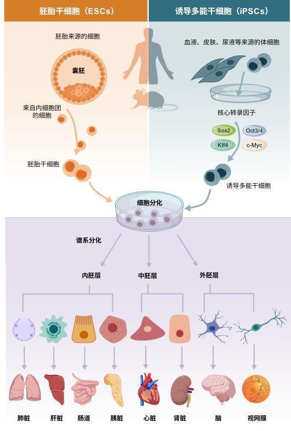
图1 多能干细胞分类及主要生物学特性[25]

注:图片使用BioGDP网站制作,下图同。

Fig. 1 Classification of pluripotent stem cells and their major biological characteristics

Note: The figure was created using BioGDP.com. Same for the following figures.

近期,研究人员相继证实,短暂激活BMP信号可高效诱导PSC向结肠类器官分化[26];而短暂激活Wnt信号则能将其定向分化为尿路上皮类器官[27]。此外,亦有研究发现,Wnt、BMP和FGF信号通路共同调控肾单位的近端和远端细胞命运,可以实现肾单位细胞命运的按需转换[28]。超越分化阶段,信号调控在类器官功能维持中同样重要,如胆酸-FXR信号的激活可有效阻止iPSC衍生肝类器官的导管化转分化,从而保障其长期培养下的肝细胞特性稳定[29]。
同时,生成类器官的分化过程也需要物理条件的支撑,细胞外基质(Extracellular matrix, ECM)是类器官3D培养的另一基石。它不仅能提供细胞黏附、增殖和组织结构的物理支架,还蕴含了复杂的生化信号[30]。目前最广泛应用的是基质胶,它源自小鼠肉瘤,富含层粘连蛋白、IV型胶原等多种ECM成分和生长因子,能有效支持类器官生长[31-32]。然而,其免疫原性、批次差异性和致病性感染的风险促使了替代方案的发展[33]。水凝胶应运而生,它具有高度可定制性、成分明确和稳定性好等优点[34]。已有研究证明,Arg-Gly-Asp(RGD)等整合素结合肽,可以接到水凝胶中并赋予其细胞黏附特性[35]。hiPSC类器官的构建已逐步向无基质胶和无支架体系发展,利用工程化ECM支架在早期阶段提供支持,随后支架降解以促进iPSC自组装。除了传统的基质胶包埋培养,类器官培养技术也在不断革新,出现了气液界面培养[36]、旋转细胞培养系统[37]、微流体技术[38]以及CellRaft AIR系统[39]等多种模式。这些新兴技术旨在更好地模拟生理环境、实现高通量筛选或满足特定的实验需求,共同推动了类器官研究领域向更精准、更可控的方向发展。

2 疾病病理机制建模

近年来,随着类器官技术的快速发展,基于iPSC和ESC衍生的各类器官模型,已成为揭示疾病发生发展机制的重要前沿平台。这些三维培养体系不仅高度模拟人体器官的关键结构与发育动态,更在转录组、表观遗传等多层次再现生理与病理状态,为深入研究神经、心脏、肝、肾及肠道等系统的疾病机制提供了前所未有的体外模型。

2.1 神经类器官

基于人诱导多能干细胞和人胚胎干细胞来源的神经类器官,因其能够高度模拟早期人脑发育的关键进程,已成为研究神经发育与疾病的重要体外模型。此类模型不仅能模拟发育中大脑的复杂结构,如顶端-基底极性分布的祖细胞区域及多样化的神经元类型,还因其转录组与表观基因组特征与人胎儿大脑高度吻合,具备良好的生理相关性[40-41]。全基因组甲基化与转录组分析显示,类器官在分化过程中能重现胎儿大脑从早期至中期的基因表达动态,其中富含非CG甲基化的基因组区域与超级增强子重叠,这些区域在胎儿期活跃转录,而在产后发育阶段被抑制;同时,类器官中也检测到与人胎儿大脑一致的转录因子结合基序富集及启动子DNA甲基化模式,充分验证了该模型在表观遗传层面的可靠性[42]。
在阿尔茨海默病研究中,研究人员利用血清暴露模拟疾病中的血脑屏障损伤,成功在类器官中重现了β-淀粉样蛋白沉积、Tau蛋白过度磷酸化、突触功能受损以及星形胶质细胞免疫激活等一系列关键病理事件[43]。在C9ORF72突变相关的肌萎缩侧索硬化/额颞叶痴呆研究中,患者来源的iPSC类器官模型不仅再现了RNA焦点、二肽重复蛋白积聚等分子病理特征,还揭示了DNA损伤应答异常及早期的神经发育紊乱[44-45]。

2.2 心脏类器官

诱导多能干细胞来源的心脏类器官已成为研究心脏疾病机制的关键模型,在疾病建模方面,研究者利用患者特异性iPSC结合CRISPR-Cas9基因校正技术,成功模拟了遗传性心脏病如扩张型心肌病与肥厚型心肌病的病理特征[46-47]。扩张型心肌病模型揭示了肌节蛋白TTN聚集导致蛋白质质量控制异常及单倍体不足等机制[48-49];肥厚型心肌病模型则证实MYBPC3突变可通过激活无义介导的mRNA降解通路致病,而MYH7突变则通过旁分泌信号激活间质细胞,促使胶原沉积和组织纤维化[50-51]。

2.3 肺类器官

多能干细胞可分化成2种类型的肺类器官模型,分别是肺类器官和芽尖前体类器官,其中肺类器官适用于模拟发育过程中上皮-间质相互作用,芽尖前体类器官则更适用于上皮细胞命运决定机制研究[52]。笔者所在团队基于单细胞测序技术解析了猪肺发育过程中的关键细胞类型与调控机制,建立了从猪多能干细胞经定形内胚层、前肠前段内胚层至肺类器官的分化体系,并证明了NKX2-1过表达可显著促进肺类器官的诱导与成熟分化[53]。

2.4 肝类器官

多能干细胞来源的肝类器官(Pluripotent stem cell-derived liver organoids, PSC-LOs)为研究遗传性和代谢性肝病提供了强大的平台,研究人员利用阿拉吉尔综合征患者的iPSCs生成肝类器官,成功再现了该病由JAG1基因突变导致的胆管细胞发育受损的表型[54]。同样,对于瓜氨酸血症1型和糖原贮积病Ia型等代谢性疾病,患者来源的PSC-LOs能够在体外重现氨积累、脂质和糖原异常蓄积等关键病理特征,并可用于测试基因校正疗法的效果[55-56]。此外,利用PSC-LOs也已成功建立了酒精性肝病的模型,在模型中可观察到氧化应激、线粒体损伤、肝细胞坏死等关键病理特征[57-58]。

2.5 肠道类器官

人类多能干细胞源肠道类器官里程碑始于2011年,Wells团队首次建立了高效的体外诱导体系,成功将人多能干细胞定向分化为具有三维结构和功能的肠道类器官[59]。人源多能干细胞衍生类器官比起单纯的上皮类器官,其修复能力更强。Helmrath研究团队证实[60],将人源多能干细胞衍生的肠道类器官移植至小鼠受损肠道后,能够有效修复包括上皮和深层组织在内的肠壁结构,并恢复小肠的功能。

2.6 肾类器官

体外肾生成过程始于人多能干细胞分化为中间中胚层细胞,随后进一步诱导为后肾间充质和输尿管芽细胞群,这两种细胞群体通过相互作用共同构建肾脏结构:后肾间充质产生肾元前体并最终形成肾单位,而输尿管芽则通过分支形态发生发育为集合管系统[21,61-62]。2015年,Freedman等[63]首次建立了将人多能干细胞分化为肾脏类器官的方案,并通过敲除PKD1或PKD2基因构建了首个常染色体显性多囊肾病类器官模型。随后在2021年,该团队利用CD24+肾上皮细胞群建立了可快速诱导囊肿形成的常染色体显性多囊肾病模型[64]。2024年,他们利用碱基编辑技术,进一步构建了能模拟4种常见无意义突变的PKD类器官,并鉴定出真核核糖体选择性氨基糖苷类药物可作为潜在的治疗药物[65]。

3 应用

类器官模型作为高度仿生的三维体外系统,已成为连接基础研究与临床转化的重要桥梁。本部分将系统介绍类器官在感染与肿瘤模型构建、药物筛选与毒性评估等方面的最新进展,重点阐述其在模拟疾病过程、揭示致病机制、开发新型疗法及评价药物安全性中的关键作用(图2)。
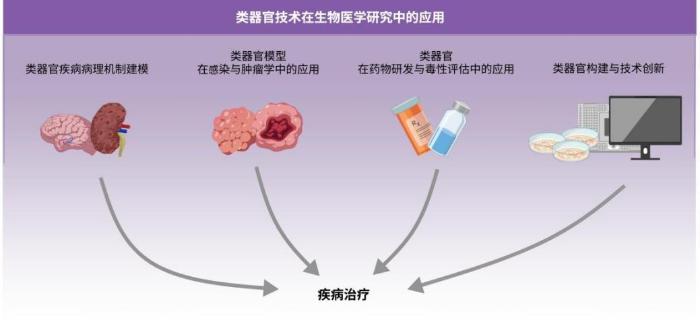
图2 类器官在疾病建模、药物筛选中的应用示意图[13]

Fig. 2 Schematic illustration of organoid applications in disease modeling and drug screening

3.1 感染与肿瘤模型应用

在寨卡病毒研究中,人类多能干细胞衍生的脑类器官揭示该病毒可导致类器官尺寸显著减小、神经祖细胞大量凋亡及有丝分裂异常,并阐明了这些病理改变与Toll样受体3激活间的联系[66-68]。这一发现不仅确立了脑类器官在病毒致畸机制研究中的重要地位,也为筛选针对特定信号通路的抗病毒药物提供了理想模型。
在严重急性呼吸综合征冠状病毒2(Severe acute respiratory syndrome coronavirus 2, SARS-CoV-2)研究中,肺类器官被证实可重现SARS-CoV-2的感染机制,并验证了多种抑制剂(如iPep-SARS2-E、E24等)的抗病毒效力[69-70]。尽管COVID-19主要导致呼吸衰竭,但其常伴有肝损伤等肝外并发症,研究团队利用PSC生成的肝脏和胰腺类器官研究SARS-CoV-2的病毒趋向性,研究证实胰腺α和β细胞以及肝脏类器官均易受SARS-CoV-2感染[71-72]。Zhang等[73]在感染后的脑类器官冷冻切片中直接观测到病毒核衣壳蛋白表达及透射电镜下的病毒颗粒,并通过免疫标记共定位证实病毒可直接感染皮质神经元与神经祖细胞,提示其对中枢神经系统的潜在侵袭能力。综上所述,多谱系类器官模型有效地揭示了SARS-CoV-2的广泛组织嗜性及潜在的全身性病理影响,为了解病毒的复杂致病机理提供了系统性视角。
戊型肝炎病毒(Hepatitis E virus, HEV)是全球急性病毒性肝炎的主要致病源,通过人iPSC来源的肝脏、肠道及脑类器官模型,首次系统证实HEV具有跨器官感染能力,肝脏与肠道类器官支持HEV1/3a/4完成完整生命周期,而脑类器官仅对HEV1/3a易感,揭示了神经系统感染的基因型特异性[74]。这些结果表明,类器官系统能够精准解析HEV的跨组织传播路径及基因型依赖的感染特征,弥补了传统细胞模型在宿主特异性研究中的不足。
在肿瘤研究领域,类器官胶质瘤模型比起传统的二维共培养,更能真实地模拟胶质瘤干细胞对替莫唑胺、双-氯乙基亚硝基脲等化疗药物及电离辐射的体内耐药性,表现为遗传毒性应激水平更低[75]。为了提高此类3D模型的筛选效率,研究人员开发了一种基于微型环状几何结构的测定系统,该系统通过在96孔板中形成环形类器官阵列,有效避免了操作过程中的结构损伤,实现了非破坏性的换液与加药,为高通量药物筛选提供了可靠平台[75]。从小鼠模型和人类iPSC衍生的祖体分离出的基因改造细胞,以及人类肺肿瘤来源细胞,都已被用于生成肺肿瘤模型。利用人类基因改造的iPSC衍生祖细胞,研究者成功构建了肺肿瘤类器官,其在形态、组织结构和遗传特征上与原生肿瘤高度相似[76-77]。总之,肿瘤类器官模型结合创新的培养工程技术,不仅高度还原了肿瘤的体内生物学特性,更为抗肿瘤药物的高效筛选与耐药机制研究提供了具有临床转化价值的实验平台。

3.2 药物研发与毒性评估

在药物毒性筛选与治疗策略开发领域,基于多电极阵列的高通量平台已成功用于评估多种药物(如多柔比星及酪氨酸激酶抑制剂)的急慢性心脏毒性,有效识别出场电位时程改变、心律失常及阻抗下降等表型[78]。此外,通过CRISPR筛选结合类器官模型,研究人员鉴定出碳酸酐酶12作为多柔比星心脏毒性的潜在干预靶点,并发现药物双硫仑可有效缓解毒性表型[79]。Lucassen等[80]将iPSC来源的内耳类器官作为耳毒性模型,发现顺铂和庆大霉素可以剂量依赖性地诱导毛细胞与神经元丢失、结构破坏及细胞凋亡。在此之前,有研究人员利用iPSC衍生的间充质干细胞外泌体作为载体封装顺铂等化疗药物,并在自体iPSC衍生的肺及肺癌类器官模型中评估其靶向递送效率,这一成果为肺癌的精准治疗开辟了新路径[81]。近期有研究团队利用人多能干细胞衍生的肝类器官模型揭示了登革病毒主要靶向增殖中的类肝细胞并引发线粒体损伤,进而通过药物筛选他们发现激活NRF2通路的氧白藜芦醇和奥马维酮能够通过维持线粒体功能有效抑制病毒,为抗登革病毒治疗提供了新策略[82]。特别值得注意的是,Greka实验室基于类器官系统的药物发现研究取得了突破性进展[83],他们发现黏蛋白1肾病患者来源肾脏类器官中累积的错误折叠的MUC1-fs蛋白被TMED9货物受体囊泡捕获,并鉴定出小分子化合物BRD4780能够通过结合TMED9释放MUC1-fs,为粘蛋白1肾病治疗提供了潜在新药。综上所述,结合了基因编辑与高通量筛选等前沿技术的类器官系统,不仅显著提升了药物安全性评估的临床预测价值,更通过精准解析致病机理与药物作用靶点,为加速新药发现及个性化治疗策略的转化应用提供了强有力的实验平台。

4 模型构建与技术创新

类器官模型的不断完善与创新,正极大地推动着其生理相关性与应用边界的拓展。为突破传统模型在血管化、细胞异质性与功能复杂性等方面的局限,一系列前沿技术应运而生。
为克服传统模型缺乏血管系统的局限性,新兴的“组装技术”将血动脉类器官(包含内皮细胞、周细胞等)与神经类器官共培养,成功构建出具有功能性毛细血管网络和血脑屏障样结构的脑血管类器官[84-85]。Anton-Bolanos等[86]开发的“脑嵌合体”模型,通过在同一大脑类器官中共同培养多供体干细胞,揭示了不同遗传背景的神经前体细胞和神经元对毒性刺激的反应存在显著差异,为个体化毒性评估与神经疾病研究提供了新型可扩展平台。此外,将人类iPSC衍生的小胶质细胞整合至神经类器官或建立共培养体系,成功构建了能够模拟神经炎症的类器官组装模型[87]。
近日,有研究通过钙成像、高密度微电极阵列等技术,证实人脑类器官具备学习和记忆所需的基础构建模块,包括突触形成、受体表达、即时早期基因激活、突触可塑性以及复杂的神经元网络动态等[88]。另外,约翰斯·霍普金斯大学科研团队成功开发出具有可编程特性的形状自适应壳装微电极阵列,实现了对三维心脏类器官的全表面电生理映射与长期动态监测,极大提升了数据采集的时空分辨率,为精准解析疾病网络与评估药物效应提供了强大工具[89]。近期,Kim等[90]模拟胚胎植入与侵袭的过程,在具有高孔隙率、高亲水性和低刚度的胶原-聚乙二醇复合凝胶上培养胚状体,获得了具有腔室结构和同步收缩功能的心脏类器官,为心血管转化研究提供了简洁有效的平台。
结合促进肝细胞成熟的中胚层衍生旁分泌信号,以及介导细胞间接触的三维微环境,肝类器官的三维培养技术逐渐成熟[91-92]。Takebe等[91]通过共培养PSC来源的肝内胚层细胞、人间充质干细胞和内皮细胞,成功生成了具有三维结构的肝芽,移植到小鼠体内后,这些肝芽能够与宿主循环系统连接并进一步成熟,证明了功能性血管化对于类器官成熟的关键作用。近日,通过在iPSC分化过程中引入维生素C和胆红素等因子,诱导出具有区域特异性的肝前体细胞进行自我组装,形成了包含三个肝区特征的类器官[93]。之后,另一研究团队通过分阶段诱导iPSC分化为红细胞-髓系祖细胞与肝内胚层,并结合内皮与间充质细胞,成功构建了包含功能库普弗细胞的肝类器官,该模型首次实现了肝细胞、内皮细胞、间充质细胞与免疫细胞的共同培养,为研究肝病机制与药物筛选提供了生理相关性更高的全新平台[94]。
Yin等[95]设计了一种立方体芯片培养平台,可以从PSC分化出具有远端呼吸气道特征的类器官,该类器官能够在30 d内实现结构成熟,用于模拟慢性阻塞性肺疾病及细胞治疗。研究团队不断优化分化方案,通过在分化体系中引入肠道干细胞微环境因子表皮调节素,成功在单一流程中同步生成了包含上皮、间质、平滑肌、功能性肠神经元及血管前体细胞的复杂类器官,显著提升了类器官的结构完整性与生理相关性[96]。随后,另一团队首次利用人诱导多能干细胞,通过模拟胚胎发育中中胚层与内胚层的协同过程,成功构建出高度血管化且具备器官特异性的肺和肠道类器官[97]。
研究人员首次成功利用干细胞在实验室培育出能够持续生长数月的人类肾脏类器官,模拟了胎儿肾脏的发育过程[98]。另一项突破性研究则利用人多能干细胞构建出具有复杂三维结构的肾脏类器官,这些类器官不仅显示出肾脏的主要生理功能,在移植后还能产生类似尿液的体液,为构建可移植人工肾脏奠定了基础,同时在疾病模拟和药物研发领域展现出重要应用前景[99]。在前期成功构建人-小鼠嵌合肾类器官的基础上,Yokoo团队优化了培养体系,使人猪两个物种的肾脏能够实现连续发育,从而成功生成了人-猪嵌合肾类器官[100-101]。近期,Noguchi等[102]发现过表达肾癌相关融合基因VCL-ALK,可诱导肾小管细胞持续增殖,且这些细胞滞留在未成熟的肾小管祖细胞阶段,成功构建了可模拟肾肿瘤发生早期阶段的体外模型。

5 总结与展望

多能干细胞与三维类器官技术的深度结合,正把体外模型研究推向真正的“类生理”的时代。从该领域的核心进展来看,研究者通过精准调控Wnt、BMP、TGF-β等发育信号通路,再配合基质胶或水凝胶这类工程化支架,可以成功诱导多能干细胞分化成脑、心、肝、肾及肠道等复杂类器官。这些体外模型不但在三维结构和基因表达上能较好地模拟了原生器官,还展现出了实质性的生理功能。脑类器官具有神经电活动,心脏类器官能够节律性地搏动,肝肾模型也具备一定的代谢与滤过能力,这些现象都证明了它们的生物学相关性。
在应用层面,多能干细胞源类器官已经不仅是基础研究的工具,而是逐渐成为转化医学中一个可用的平台。它们不仅能够重现遗传性疾病的早期病理变化,还能模拟病毒的细胞特异性感染和损伤机制,同时在肿瘤微环境建模和解析药物耐药性方面,都展现出了比传统模型更好的功能。
尽管多能干细胞源类器官技术前景较好,但在畜禽种业中的应用仍面临不少挑战。一是细胞来源的问题,畜禽多能干细胞在体外诱导分化方面技术门槛比较高,所以目前相关研究远少于畜禽成体干细胞源类器官领域,但是多能干细胞具备无限增殖的能力,也能做精准的基因编辑,从长远角度看是实现高通量育种的合适载体。二是标准化和质量控制,需要建立一套统一的培养和质控体系,开发成分明确、可以合成的仿生基质,逐步降低对动物来源基质胶的依赖,从而 减少现有培养中的批次差异和细胞异质性等问题。三是把这项技术真正推向育种实践,通过大规模群体验证多能干细胞源类器官在抗病性评估、生长性能预测等方面的可靠性,同时建立一套适合畜禽育种应用的评价与监管体系。
综上所述,多能干细胞源类器官技术在畜禽领域正处在一个从技术突破逐步走向育种应用的关键节点。虽然目前畜禽多能干细胞源类器官研究还处于起步阶段,但人源类器官在复杂病理模型中的成熟应用已经提供了不少可参考的经验。展望未来,随着持续优化培养体系、推动跨学科技术融合,畜禽多能干细胞源类器官有望解决实验周期长、成本高等问题,从而提升畜禽的抗病育种效率,加快优良性状的筛选,最终为畜禽种业的创新和可持续发展提供新的技术支撑。
[1]
EVANS M J, KAUFMAN M H. Establishment in culture of pluripotential cells from mouse embryos[J]. Nature, 1981, 292(5819): 154-156.

[2]
THOMSON J A, ITSKOVITZ-ELDOR J, SHAPIRO S S, et al. Embryonic stem cell lines derived from human blastocysts[J]. Science, 1998, 282(5391): 1145-1147.

[3]
VOLAREVIC V, MARKOVIC B S, GAZDIC M, et al. Ethical and safety issues of stem cell-based therapy[J]. International Journal of Medical Sciences, 2018, 15(1): 36-45.

[4]
TAKAHASHI K, TANABE K, OHNUKI M, et al. Induction of pluripotent stem cells from adult human fibroblasts by defined factors[J]. Cell, 2007, 131(5): 861-872.

[5]
PARK I H, ZHAO R, WEST J A, et al. Reprogramming of human somatic cells to pluripotency with defined factors[J]. Nature, 2008, 451(7175): 141-146.

[6]
YU J Y, VODYANIK M A, SMUGA-OTTO K, et al. Induced pluripotent stem cell lines derived from human somatic cells[J]. Science, 2007, 318(5858): 1917-1920.

[7]
LIU Y Y, WU D K, CHEN J B, et al. Advances in the study of gastric organoids as disease models[J]. World Journal of Gastrointestinal Oncology, 2024, 16(5): 1725-1736.

[8]
KRETZSCHMAR K, CLEVERS H. Organoids: modeling development and the stem cell niche in a dish[J]. Developmental Cell, 2016, 38(6): 590-600.

[9]
SATO T, VRIES R G, SNIPPERT H J, et al. Single Lgr5 stem cells build crypt-villus structures in vitro without a mesenchymal niche[J]. Nature, 2009, 459(7244): 262-265.

[10]
CAMP J G, BADSHA F, FLORIO M, et al. Human cerebral organoids recapitulate gene expression programs of fetal neocortex development[J]. Proceedings of the National Academy of Sciences of the United States of America, 2015, 112(51): 15672-15677.

[11]
ZHOU T, TAN L, CEDERQUIST G Y, et al. High-content screening in hPSC-neural progenitors identifies drug candidates that inhibit zika virus infection in fetal-like organoids and adult brain[J]. Cell Stem Cell, 2017, 21(2): 274-283.e5.

[12]
KIM J, KOO B K, KNOBLICH J A. Human organoids: model systems for human biology and medicine[J]. Nature Reviews Molecular Cell Biology, 2020, 21(10): 571-584.

[13]
EIRAKU M, WATANABE K, MATSUO-TAKASAKI M, et al. Self-organized formation of polarized cortical tissues from ESCs and its active manipulation by extrinsic signals[J]. Cell Stem Cell, 2008, 3(5): 519-532.

[14]
LANCASTER M A, KNOBLICH J A. Organogenesis in a dish: modeling development and disease using organoid technologies[J]. Science, 2014, 345(6194): 1247125.

[15]
CHAMBERS S M, FASANO C A, PAPAPETROU E P, et al. Highly efficient neural conversion of human ES and iPS cells by dual inhibition of SMAD signaling[J]. Nature Biotechnology, 2009, 27(3): 275-280.

[16]
EIRAKU M, TAKATA N, ISHIBASHI H, et al. Self-organizing optic-cup morphogenesis in three-dimensional culture[J]. Nature, 2011, 472(7341): 51-56.

[17]
WAHLIN K J, MARUOTTI J A, SRIPATHI S R, et al. Photoreceptor outer segment-like structures in long-term 3D retinas from human pluripotent stem cells[J]. Scientific Reports, 2017, 7: 766.

[18]
LACO F, LOW J L, SEOW J, et al. Cardiomyocyte differentiation of pluripotent stem cells with SB203580 analogues correlates with Wnt pathway CK1 inhibition independent of p38 MAPK signaling[J]. Journal of Molecular and Cellular Cardiology, 2015, 80: 56-70.

[19]
LIAN X J, HSIAO C, WILSON G, et al. Robust cardiomyocyte differentiation from human pluripotent stem cells via temporal modulation of canonical Wnt signaling[J]. Proceedings of the National Academy of Sciences of the United States of America, 2012, 109(27): E1848-E1857.

[20]
TAGUCHI A, KAKU Y, OHMORI T, et al. Redefining the in vivo origin of metanephric nephron progenitors enables generation of complex kidney structures from pluripotent stem cells[J]. Cell Stem Cell, 2014, 14(1): 53-67.

[21]
ANG L T, TAN A K Y, AUTIO M I, et al. A roadmap for human liver differentiation from pluripotent stem cells[J]. Cell Reports, 2018, 22(8): 2190-2205.

[22]
D’AMOUR K A, BANG A G, ELIAZER S, et al. Production of pancreatic hormone–expressing endocrine cells from human embryonic stem cells[J]. Nature Biotechnology, 2006, 24(11): 1392-1401.

[23]
DYE B R, HILL D R, FERGUSON M A, et al. In vitro generation of human pluripotent stem cell derived lung organoids[J]. eLife, 2015, 4: e05098.

[24]
MCCRACKEN K W, HOWELL J C, WELLS J M, et al. Generating human intestinal tissue from pluripotent stem cells in vitro [J]. Nature Protocols, 2011, 6(12): 1920-1928.

[25]
JIANG S, LI H Q, ZHANG L, et al. Generic Diagramming Platform (GDP): a comprehensive database of high-quality biomedical graphics[J]. Nucleic Acids Research, 2025, 53(D1): D1670-D1676.

[26]
MÚNERA J O, SUNDARAM N, RANKIN S A, et al. Differentiation of human pluripotent stem cells into colonic organoids via transient activation of BMP signaling[J]. Cell Stem Cell, 2017, 21(1): 51-64.e6.

[27]
QU N, DAOUD A, KECHELE D O, et al. Differentiation of human pluripotent stem cells into urothelial organoids via transient activation of WNT signaling[J]. iScience, 2025, 28(5): 112398.

[28]
ACHIENG M A, SCHNELL J, FAUSTO C C, et al. Axial nephron fate switching demonstrates a plastic system tunable on demand[J]. Nature Communications, 2025, 16: 7912.

[29]
SHIMIZU T, MIYOSHI M, KAKINUMA S, et al. Bile acid-FXR signaling facilitates the long-term maintenance of hepatic characteristics in human iPSC-derived organoids[J]. Cell Reports, 2025, 44(5): 115675.

[30]
BONNANS C, CHOU J, WERB Z. Remodelling the extracellular matrix in development and disease[J]. Nature Reviews Molecular Cell Biology, 2014, 15(12): 786-801.

[31]
ASANO Y, NISHIGUCHI A, MATSUSAKI M, et al. Ultrastructure of blood and lymphatic vascular networks in three-dimensional cultured tissues fabricated by extracellular matrix nanofilm-based cell accumulation technique[J]. Mic-roscopy, 2014, 63(3): 219-226.

[32]
ISAEVA E V, BEKETOV E E, ARGUCHINSKAYA N V, et al. Decellularized extracellular matrix for tissue engineering (review)[J]. Sovremennye Tehnologii v Medicine, 2022, 14(3): 57.

[33]
WANG Y L, GUNASEKARA D B, REED M I, et al. A microengineered collagen scaffold for generating a polarized crypt-villus architecture of human small intestinal epithelium[J]. Biomaterials, 2017, 128: 44-55.

[34]
ZACHOS N C, KOVBASNJUK O, FOULKE-ABEL J, et al. Human enteroids/colonoids and intestinal organoids functionally recapitulate normal intestinal physiology and pathophysiology[J]. Journal of Biological Chemistry, 2016, 291(8): 3759-3766.

[35]
BLONDEL D, LUTOLF M P. Bioinspired hydrogels for 3D organoid culture[J]. Chimia, 2019, 73(1/2): 81.

[36]
GIANDOMENICO S L, MIERAU S B, GIBBONS G M, et al. Cerebral organoids at the air–liquid interface generate diverse nerve tracts with functional output[J]. Nature Neuroscience, 2019, 22(4): 669-679.

[37]
KUMAR S, JACH D, MACFARLANE W, et al. A 3-dimensional coculture model to visualize and monitor interaction between pancreatic cancer and islet β cells[J]. Pancreas, 2021, 50(7): 982-989.

[38]
HUANG J J, XU Z Y, JIAO J, et al. Microfluidic intestinal organoid-on-a-chip uncovers therapeutic targets by reca-pitulating oxygen dynamics of intestinal IR injury[J]. Bioactive Materials, 2023, 30: 1-14.

[39]
STERN A, THOMPSON B, WILLIAMS K, et al. The CellRaft AIRⓇ system: a novel system enabling organoid imaging, identification, and isolation[J]. SLAS Discovery, 2022, 27(3): 201-208.

[40]
LANCASTER M A, RENNER M, MARTIN C A, et al. Cerebral organoids model human brain development and microcephaly[J]. Nature, 2013, 501(7467): 373-379.

[41]
QIAN X Y, NGUYEN H N, SONG M M, et al. Brain-region-specific organoids using mini-bioreactors for modeling ZIKV exposure[J]. Cell, 2016, 165(5): 1238-1254.

[42]
LUO C Y, LANCASTER M A, CASTANON R, et al. Cerebral organoids recapitulate epigenomic signatures of the human fetal brain[J]. Cell Reports, 2016, 17(12): 3369-3384.

[43]
CHEN X W, SUN G Q, TIAN E, et al. Modeling sporadic Alzheimer's disease in human brain organoids under serum exposure[J]. Advanced Science, 2021, 8(18): 2101462.

[44]
SZEBÉNYI K, WENGER L M D, SUN Y, et al. Human ALS/FTD brain organoid slice cultures display distinct early astrocyte and targetable neuronal pathology[J]. Nature Neu-roscience, 2021, 24(11): 1542-1554.

[45]
VAN DER GEEST A T, JAKOBS C E, LJUBIKJ T, et al. Molecular pathology, developmental changes and synaptic dysfunction in (pre-) symptomatic human C9ORF72-ALS/FTD cerebral organoids[J]. Acta Neuropathologica Communications, 2024, 12(1): 152.

[46]
ELLIS B W, RONAN G, REN X, et al. Human heart Anoxia and reperfusion tissue (HEART) model for the rapid study of exosome bound miRNA expression as biomarkers for myocardial infarction[J]. Small, 2022, 18(28): 2201330.

[47]
EWOLDT J K, DEPALMA S J, JEWETT M E, et al. Induced pluripotent stem cell-derived cardiomyocyte in vitro models: benchmarking progress and ongoing challenges[J]. Nature Methods, 2025, 22(1): 24-40.

[48]
FOMIN A, GÄRTNER A, CYGANEK L, et al. Truncated titin proteins and titin haploinsufficiency are targets for functional recovery in human cardiomyopathy due to TTN mutations[J]. Science Translational Medicine, 2021, 13(618): eabd3079.

[49]
HUANG G Y, BISARIA A, WAKEFIELD D L, et al. Titin-truncating variants in hiPSC cardiomyocytes induce pathogenic proteinopathy and sarcomere defects with preserved core contractile machinery[J]. Stem Cell Reports, 2023, 18(1): 220-236.

[50]
SEEGER T, SHRESTHA R, LAM C K, et al. A premature termination codon mutation in MYBPC3 causes hypertrophic cardiomyopathy via chronic activation of nonsense-mediated decay[J]. Circulation, 2019, 139(6): 799-811.

[51]
EWOLDT J K, WANG M C, MCLELLAN M A, et al. Hypertrophic cardiomyopathy-associated mutations drive stro-mal activation via EGFR-mediated paracrine signaling[J]. Science Advances, 2024, 10(42): eadi6927.

[52]
MILLER A J, DYE B R, FERRER-TORRES D, et al. Generation of lung organoids from human pluripotent stem cells in vitro [J]. Nature Protocols, 2019, 14(2): 518-540.

[53]
YANG X C, WU X L, WANG Y Q, et al. Induction of lung progenitor cell-like organoids by porcine pluripotent stem cells[J]. The FASEB Journal, 2024, 38(4): e23481.

[54]
GUAN Y, XU D, GARFIN P M, et al. Human hepatic organoids for the analysis of human genetic diseases[J]. JCI Insight, 2017, 2(17): e94954.

[55]
AKBARI S, SEVINÇ G G, ERSOY N, et al. Robust, long-term culture of endoderm-derived hepatic organoids for disease modeling[J]. Stem Cell Reports, 2019, 13(4): 627-641.

[56]
MUN S J, HONG Y H, SHIN Y, et al. Efficient and reproducible generation of human induced pluripotent stem cell-derived expandable liver organoids for disease modeling[J]. Scientific Reports, 2023, 13: 22935.

[57]
WANG S Y, WANG X, TAN Z L, et al. Human ESC-derived expandable hepatic organoids enable therapeutic liver repo-pulation and pathophysiological modeling of alcoholic liver injury[J]. Cell Research, 2019, 29(12): 1009-1026.

[58]
FENG Z W, ZHOU B R, SHUAI Q Z, et al. Development of an alcoholic liver disease model for drug evaluation from human induced pluripotent stem cell-derived liver organoids[J]. Acta Biochimica et Biophysica Sinica, 2024, 56(10): 1460-1472.

[59]
SPENCE J R, MAYHEW C N, RANKIN S A, et al. Directed differentiation of human pluripotent stem cells into intestinal tissue in vitro [J]. Nature, 2011, 470(7332): 105-109.

[60]
POLING H M, SUNDARAM N, FISHER G W, et al. Human pluripotent stem cell-derived organoids repair damaged bowel in vivo [J]. Cell Stem Cell, 2024, 31(10): 1513-1523.

[61]
TAKASATO M, ER P X, CHIU H S, et al. Generation of kidney organoids from human pluripotent stem cells[J]. Nature Pro-tocols, 2016, 11(9): 1681-1692.

[62]
TAKASATO M, ER P X, CHIU H S, et al. Kidney organoids from human iPS cells contain multiple lineages and model human nephrogenesis[J]. Nature, 2015, 526(7574): 564-568.

[63]
FREEDMAN B S, BROOKS C R, LAM A Q, et al. Modelling kidney disease with CRISPR-mutant kidney organoids derived from human pluripotent epiblast spheroids[J]. Nature Com-munications, 2015, 6: 8715.

[64]
XU Y X, KUPPE C, PERALES-PATÓN J, et al. Adult human kidney organoids originate from CD24+ cells and represent an advanced model for adult polycystic kidney disease[J]. Nature Genetics, 2022, 54(11): 1690-1701.

[65]
VISHY C E, THOMAS C, VINCENT T, et al. Genetics of cystogenesis in base-edited human organoids reveal therapeutic strategies for polycystic kidney disease[J]. Cell Stem Cell, 2024, 31(4): 537-553.

[66]
DANG J, TIWARI S K, LICHINCHI G, et al. Zika virus depletes neural progenitors in human cerebral organoids through activation of the innate immune receptor TLR3[J]. Cell Stem Cell, 2016, 19(2): 258-265.

[67]
GARCEZ P P, LOIOLA E C, MADEIRO DA COSTA R, et al. Zika virus impairs growth in human neurospheres and brain organoids[J]. Science, 2016, 352(6287): 816-818.

[68]
GABRIEL E, RAMANI A, KAROW U, et al. Recent zika virus isolates induce premature differentiation of neural progenitors in human brain organoids[J]. Cell Stem Cell, 2017, 20(3): 397-406.

[69]
SALAHUDEEN A A, CHOI S S, RUSTAGI A, et al. Progenitor identification and SARS-CoV-2 infection in human distal lung organoids[J]. Nature, 2020, 588(7839): 670-675.

[70]
BEKDASH R, YOSHIDA K, NAIR M S, et al. Developing inhibitory peptides against SARS-CoV-2 envelope protein[J]. PLoS Biology, 2024, 22(3): e3002522.

[71]
YANG L L, HAN Y L, NILSSON-PAYANT B E, et al. A human pluripotent stem cell-based platform to study SARS-CoV-2 tropism and model virus infection in human cells and organoids[J]. Cell Stem Cell, 2020, 27(1): 125-136.

[72]
ZHU L H, SHE Z G, CHENG X, et al. Association of blood glucose control and outcomes in patients with COVID-19 and pre-existing type 2 diabetes[J]. Cell Metabolism, 2020, 31(6): 1068-1077.

[73]
ZHANG B Z, CHU H, HAN S, et al. SARS-CoV-2 infects human neural progenitor cells and brain organoids[J]. Cell Research, 2020, 30(10): 928-931.

[74]
LIU F, LIU T X, WU X, et al. iPSC-induced multilineage liver organoids, small intestinal organoids and brain organoids sustain pangenotype hepatitis E virus propagation[J]. Gut, 2026, 75(4): 787-800.

[75]
LINKOUS A, BALAMATSIAS D, SNUDERL M, et al. Modeling patient-derived glioblastoma with cerebral organoids[J]. Cell Reports, 2019, 26(12): 3203-3211.

[76]
KIM M, MUN H, SUNG C O, et al. Patient-derived lung cancer organoids as in vitro cancer models for therapeutic screening[J]. Nature Communications, 2019, 10: 3991.

[77]
YOKOTA E, IWAI M, YUKAWA T, et al. Clinical application of a lung cancer organoid (tumoroid) culture system[J]. npj Precision Oncology, 2021, 5: 29.

[78]
ALTROCCHI C, VAN AMMEL K, STEEMANS M, et al. Evaluation of chronic drug-induced electrophysiological and cytotoxic effects using human-induced pluripotent stem cell-derived cardiomyocytes (hiPSC-CMs)[J]. Frontiers in Pha-rmacology, 2023, 14: 1229960.

[79]
LIU C, SHEN M C, LIU Y X, et al. CRISPRi/a screens in human iPSC-cardiomyocytes identify glycolytic activation as a druggable target for doxorubicin-induced cardiotoxicity[J]. Cell Stem Cell, 2024, 31(12): 1760-1776.e9.

[80]
LUCASSEN A W A, VAN DEN BOOGAARD W M C, FOUSERT E, et al. Human induced pluripotent stem cell-derived inner ear organoids reveal hair cell damage and plasticity after cisplatin and gentamicin exposure[J]. Disease Models & Mechanisms, 2026, 19(6): dmm052511.

[81]
KÜSTERMANN C, NARBUTE K, MOVČANA V, et al. iPSC-derived lung and lung cancer organoid model to evaluate cisplatin encapsulated autologous iPSC-derived mesenchymal stromal cell-isolated extracellular vesicles[J]. Stem Cell Research & Therapy, 2024, 15(1): 246.

[82]
LI M Q, XU Y P, LI K, et al. Recapitulating dengue virus infection with human pluripotent stem cell-derived liver organoids for antiviral screening[J]. Nature Communications, 2025, 16: 8069.

[83]
SUBRAMANIAN A, SIDHOM E H, EMANI M, et al. Single cell census of human kidney organoids shows reproducibility and diminished off-target cells after transplantation[J]. Nature Communications, 2019, 10: 5462.

[84]
AHN Y, AN J H, YANG H J, et al. Human blood vessel organoids penetrate human cerebral organoids and form a vessel-like system[J]. Cells, 2021, 10(8): 2036.

[85]
DAO L, YOU Z, LU L, et al. Modeling blood-brain barrier formation and cerebral cavernous malformations in human PSC-derived organoids[J]. Cell Stem Cell, 2024, 31(6): 818-833.

[86]
ANTÓN-BOLAÑOS N, FARAVELLI I, FAITS T, et al. Brain Chimeroids reveal individual susceptibility to neurotoxic triggers[J]. Nature, 2024, 631(8019): 142-149.

[87]
SHIMA S, KONDO T, INOUE H. iPSC-derived neural organoids in dementia research: recent advances and future directions[J]. Neuroscience Research, 2025, 221: 104980.

[88]
HONGEN T, SAKAI K T, ITO T, et al. Human-induced pluripotent stem cell-derived neural organoids as a novel in vitro platform for developmental neurotoxicity assessment[J]. International Journal of Molecular Sciences, 2024, 25(23): 12523.

[89]
CHOI S J, LIU Z Y, YANG F Y, et al. 3D spatiotemporal electrophysiology of cardiac organoids using shell micro-electrode arrays[J]. Advanced Materials, 2026, 38(2): e06793.

[90]
KIM E M, AHN Y, WANG J, et al. Matrix-guided embryo-like invasion enables 3D heart organoids with atrioventricular synchrony-like contraction[J]. Biomaterials, 2026, 331: 124131.

[91]
TAKEBE T, SEKINE K, ENOMURA M, et al. Vascularized and functional human liver from an iPSC-derived organ bud transplant[J]. Nature, 2013, 499(7459): 481-484.

[92]
ASAI A, AIHARA E, WATSON C, et al. Paracrine signals regulate human liver organoid maturation from induced pluripotent stem cells[J]. Development, 2017, 144(6): 1056-1064.

[93]
REZA H AL, SANTANGELO C, IWASAWA K, et al. Multi-zonal liver organoids from human pluripotent stem cells[J]. Nature, 2025, 641(8065): 1258-1267.

[94]
LI Y, NIE Y Z, TANIGUCHI H. Protocol for generating liver organoids containing Kupffer cells using human iPSCs[J]. STAR Protocols, 2025, 6(4): 104138.

[95]
YIN J X, BI Z R, DAI T K, et al. Robust generation of distal respiratory airway organoids by an engineered cuboid chip[J]. Journal of Nanobiotechnology, 2026: 1-42.

[96]
CHILDS C J, POLING H M, CHEN K, et al. Coordinated differentiation of human intestinal organoids with functional enteric neurons and vasculature[J]. Cell Stem Cell, 2025, 32(4): 640-651.

[97]
MIAO Y F, PEK N M, TAN C, et al. Co-development of mesoderm and endoderm enables organotypic vascularization in lung and gut organoids[J]. Cell, 2025, 188(16): 4295-4313.

[98]
NAMESTNIKOV M, COHEN-ZONTAG O, OMER D, et al. Human fetal kidney organoids model early human nephrogenesis and Notch-driven cell fate[J]. The EMBO Journal, 2025, 44(17): 4681-4719.

[99]
HUANG B, MEDINA P, HE J C, et al. Spatially patterned kidney assembloids recapitulate progenitor self-assembly and enable high-fidelity in vivo disease modeling[J]. Cell Stem Cell, 2025, 32(10): 1614-1633.e13.

[100]
MATSUMOTO N, YAMANAKA S, MORIMOTO K, et al. Evaluation of the ability of human induced nephron progenitor cells to form chimeric renal organoids using mouse embryonic renal progenitor cells[J]. Biochemical and Biophysical Research Communications, 2023, 662: 18-25.

[101]
FUJIMORI K, YAMANAKA S, SHIMADA K, et al. Generation of human-pig chimeric renal organoids using iPSC technology[J]. Communications Biology, 2024, 7: 1278.

[102]
NOGUCHI G, BABA M, OHTAKE S, et al. Genetically engineered human iPS cell-derived kidney organoid recapi-tulates an early stage of ALK fusion gene-associated renal tumorigenesis[J]. Molecular Cancer Research: MCR, 2025: 221-231.

Outlines

/

〈 〉